Friday, September 23, 2011

Probióticos y otras bacterias


Probiotics are live microorganisms thought to be beneficial to the host organism. According to the currently adopted definition by FAO/WHO, probiotics are: "Live microorganisms which when administered in adequate amounts confer a health benefit on the host".[1] Lactic acid bacteria (LAB) and bifidobacteria are the most common types of microbes used as probiotics; but certain yeasts and bacilli may also be helpful. Probiotics are commonly consumed as part of fermented foods with specially added active live cultures; such as in yogurt, soy yogurt, or as dietary supplements.
Etymologically, the term appears to be a composite of the Latin preposition pro ("for") and the Greek adjective βιωτικός (biotic), the latter deriving from the noun βίος (bios, "life").[2]
At the start of the 20th century, probiotics were thought to beneficially affect the host by improving its intestinal microbial balance, thus inhibiting pathogens and toxin producing bacteria.[3] Today, specific health effects are being investigated and documented including alleviation of chronic intestinal inflammatory diseases,[4] prevention and treatment of pathogen-induced diarrhea,[5] urogenital infections,[6] and atopic diseases.[7]
To date, the European Food Safety Authority has rejected most claims that are made about probiotic products, saying they are unproven.[8]
Contents
===============   ===============
The lactic acid bacteria (LAB) comprise a clade of Gram-positive, low-GC, acid-tolerant, generally non-sporulating, non-respiring rod or cocci that are associated by their common metabolic and physiological characteristics. These bacteria, usually found in decomposing plants and lactic products, produce lactic acid as the major metabolic end-product of carbohydrate fermentation. This trait has, throughout history, linked LAB with food fermentations, as acidification inhibits the growth of spoilage agents. Proteinaceous bacteriocins are produced by several LAB strains and provide an additional hurdle for spoilage and pathogenic microorganisms. Furthermore, lactic acid and other metabolic products contribute to the organoleptic and textural profile of a food item. The industrial importance of the LAB is further evinced by their generally recognized as safe (GRAS) status, due to their ubiquitous appearance in food and their contribution to the healthy microflora of human mucosal surfaces. The genera that comprise the LAB are at its core Lactobacillus, Leuconostoc, Pediococcus, Lactococcus, and Streptococcus as well as the more peripheral Aerococcus, Carnobacterium, Enterococcus, Oenococcus, Sporolactobacillus, Tetragenococcus, Vagococcus, and Weisella; these belong to the order Lactobacillales.
===========   ============
Bifidobacterium is a genus of Gram-positive, non-motile, often branched anaerobic bacteria. They are ubiquitous, endosymbiotic inhabitants of the gastrointestinal tract, vagina[1][2] and mouth (B. dentium) of mammals and other animals. Bifidobacteria are one of the major genera of bacteria that make up the colon flora in mammals. Some bifidobacteria are used as probiotics.
Before the 1960s, Bifidobacterium species were collectively referred to as "Lactobacillus bifidus".
===============   ===============
Mutaflor
Mutaflor is a probiotic consisting of a viable non-pathogenic bacteria strain named Escherichia coli Nissle 1917.[1] "The Escherichia coli strain Nissle 1917-designated DSM 6601 in the German Collection for Microorganisms in Brauschweig is one of the best-examined and therapeutically relevant bacterial strains worldwide" as claimed by the manufacturer[2]
-----------------------
Actimel
Standard Actimel (excludes variations such as Actimel Light) contains:
--------------------
Lactobacillus delbrueckii subspecies bulgaricus (until 1984 known as Lactobacillus bulgaricus) is one of several bacteria used for the production of yoghurt. It is also found in other naturally fermented products. First identified in 1905 by the Bulgarian doctor Stamen Grigorov, the bacterium feeds on lactose to produce lactic acid, which is used to preserve milk.
It is a Gram-positive rod that may appear long and filamentous. It is also non-motile, and it does not form spores. This bacterium is regarded as aciduric or acidophilic, since it requires a low pH (around 5.4-4.6) to grow effectively. The bacterium has complex nutritional requirements, including the inability to ferment any sugar except lactose.
----------------
Streptococcus salivarius subsp. thermophilus (common name Streptococcus thermophilus) is a Gram-positive bacteria and a homofermentative facultative anaerobe, of the viridans group.[1] It tests negative for cytochrome, oxidase and catalase, and positive for alpha-hemolytic activity.[1] It is non-motile and does not form endospores.[1]
It is also classified as a lactic acid bacterium.[2] S. thermophilus is found in fermented milk products. It is not a probiotic (it does not survive the stomach in healthy humans) and is generally used in the production of yogurt,[3] alongside Lactobacillus bulgaricus. The two species are synergistic, and S. thermophilus probably provides L. bulgaricus with folic acid and formic acid which it uses for purine synthesis

Friday, July 15, 2011

Non-steroidal anti-inflammatory drug

Nonsteroidal anti-inflammatory drugs, usually abbreviated to NSAIDs or NAIDs, but also referred to as nonsteroidal anti-inflammatory agents/analgesics (NSAIAs) or nonsteroidal Anti-inflammatory medicines (NSAIMs), are drugs with analgesic and antipyretic (fever-reducing) effects and which have, in higher doses, anti-inflammatory effects.
The term "nonsteroidal" is used to distinguish these drugs from steroids, which, among a broad range of other effects, have a similar eicosanoid-depressing, anti-inflammatory action. As analgesics, NSAIDs are unusual in that they are non-narcotic.
The most prominent members of this group of drugs are aspirin, ibuprofen, and naproxen, all of which are available over the counter in many areas.[1][2]
Contents

Drug allergies

A drug allergy is an allergy to a drug, most commonly a medication. Medical attention should be sought immediately if an allergic reaction is suspected.
An allergic reaction will not occur on the first exposure to a substance. The first exposure allows the body to create antibodies and memory lymphocyte cells for the antigen. However, drugs often contain many different substances, including dyes, which could cause allergic reactions. This can cause an allergic reaction on the first administration of a drug. For example, a person who developed an allergy to a red dye will be allergic to any new drug which contains that red dye.
A drug allergy is different from an intolerance. A drug intolerance, which is often a milder, non-immune-mediated reaction, does not depend on prior exposure. Most people who believe they are allergic to aspirin are actually suffering from a drug intolerance.
Contents

Acne

Methicillin-resistant Staphylococcus aureus

Methicillin-resistant Staphylococcus aureus (MRSA) is a bacterium responsible for several difficult-to-treat infections in humans. It may also be called multidrug-resistant Staphylococcus aureus or oxacillin-resistant Staphylococcus aureus (ORSA).
MRSA is, by definition, any strain of Staphylococcus aureus that has developed resistance to beta-lactam antibiotics which include the penicillins (methicillin, dicloxacillin, nafcillin, oxacillin, etc.) and the cephalosporins.
MRSA is especially troublesome in hospitals and nursing homes where patients with open wounds, invasive devices and weakened immune systems are at greater risk of infection than the general public.

Chlorhexidine

Chlorhexidine is a chemical antiseptic.[1] It is effective on both Gram-positive and Gram-negative bacteria, although it is less effective with some Gram-negative bacteria.[2] It has both bactericidal and bacteriostatic mechanisms of action, the mechanism of action being membrane disruption, not ATPase inactivation as previously thought.[3] It is also useful against fungi and enveloped viruses, though this has not been extensively investigated. Chlorhexidine is harmful in high concentrations, but is used safely in low concentrations in many products, such as mouthwash and contact lens solutions.
Contents

Toxoplasmosis

Toxoplasma gondii es una especie de protozoo parásito causante de la toxoplasmosis, una enfermedad en general leve, pero que puede complicarse hasta convertirse en fatal, especialmente en los gatos y en los fetos humanos.1 El gato es su hospedador definitivo, aunque otros animales homeotermos como los humanos también pueden hospedarlo. 
Fuentes de infección
La fuente de infección más frecuente no son los animales de compañía como erróneamente se cree y se sigue difundiendo sin base científica.9
La realidad es que la fuente por la cual entra el parásito en los humanos con mayor frecuencia es a través de los alimentos contaminados: la carne (cuando está poco cocinada, ya que un gran porcentaje está contaminada) y las frutas y verduras mal lavadas.10
De las carnes disponibles para consumo en el mercado (o las carnes de caza), un gran porcentaje de todas las especies está contaminado, así que cualquier persona que consume carne ha consumido (de hecho) carne contaminada por el parásito. También es posible que por la manipulación de la carne contaminada con las manos, al llevarlas a la boca, se ingiera el parásito.
Por otro lado, una persona que consume con la necesaria frecuencia verduras y frutas, puede consumirlas sin el adecuado lavado para eliminar el parásito en algún momento. También puede consumidas manipuladas por terceros sin poder supervisar si el lavado es suficiente (por ejemplo, en restaurantes).
El ciclo vital de Toxoplasma tiene como huésped definitivo al gato o miembros de su familia, que tras ingerir alguna de las formas del parásito sufre en las células epiteliales de su intestino un ciclo asexual y luego un ciclo sexual, eliminándose en sus heces millones de ooquistes. Cuando estos esporulan se vuelven infecciosos pudiéndose infectar otros animales por su ingestión. Por debajo de 4 °C, o por encima de 37 °C, no se produce la esporulación y los quistes no son infecciosos.
Los humanos sufren la transmisión del parásito fundamentalmente por vía oral a través de la ingesta de carnes, verduras, el agua, huevos, leche, u otros alimentos contaminados por ooquistes o que contienen quistes tisulares. De hecho, hasta un 25% de las muestras de carnes de cordero y cerdo presentan ooquistes, siendo menos frecuentes en la carne de vaca. Los gatos, sobre todo si se manipulan sus excreciones, pueden infectar al ingerir los ooquistes por las manos contaminadas.

La última vía de contagio suele producirse entre personas que trabajan la tierra con las manos, bien agricultores, bien en labores de jardinería. En los suelos suele estar presente el parásito en gran cantidad. Una persona que manipule la tierra con las manos desnudas puede introducir restos de tierra bajo las uñas. Pese a un lavado de manos con agua y jabón, siempre puede quedar tierra bajo las uñas. Después, si se lleva las manos a la boca, es fácil infectarse de éste y/o de otros parásitos. Si es una persona que trabaja en el campo, no tiene por qué lavarse las manos cada vez que manipula esa tierra y en un descuido (o por mala costumbre) puede llevarse las manos sin lavar a la boca.
Siempre se ha relacionado erróneamente al gato doméstico como fuente de infección, puesto que sí son los hospedadores definitivos junto con otras especies de felinos. El error se basa en que el comportamiento humano necesario para esta infección no es el "normal".
Para que un gato pueda producir heces infecciosas tiene que contagiarse. Es decir, un gato que no está infectado y vive en una casa sin acceso al exterior y comiendo pienso o carne cocinada, no puede infectarse y por tanto no puede infectar a otros.
Si el gato tiene acceso al exterior o es silvestre, o come carne cruda, o caza pájaros o ratones y se los come, entonces sí puede infectarse.
Una vez infectado, incuba el parásito durante un periodo de entre 3 y 20 días (según la forma en la que lo ingiere, que determina la fase en la que se encuentra el parásito). Después y durante sólo un periodo de 1 mes, libera los ooquistes en las heces. Después de eso, aunque se vuelva a infectar, nunca más liberará ooquistes.
Para que esas heces con ooquistes (oocitos) sean a su vez infecciosas, necesitan un tiempo de exposición al medio de entre 24 y 48 horas. Las personas normales que conviven con gatos en casa suelen retirar las heces de los areneros con más frecuencia, impidiendo que esos ooquistes maduren y sean infecciosos. Y después, es necesario un contacto muy íntimo con esas heces para infectarse a partir de ellas. Es necesario comerse las heces del gato para infectarse (cosa que sólo hacen los niños o personas con enfermedades mentales) o si no, manipularlas con las manos y sin guardar unas mínimas medidas de higiene, llevárselas a la boca. De nuevo citamos a la "gente normal" que si tiene que realizar una limpieza de heces, de gato o de cualquier animal, después procura lavarse las manos con agua y jabón. No sólo se puede introducir el Toxoplasma Gondii en el organismo de esta manera, también otros parásitos, bacterias y virus, mucho más peligrosos e incluso letales en ocasiones como la Escherichia coli.
Por tanto, cualquier persona que conviva con un gato o varios como mascotas, incluso con acceso al exterior y hasta que coman a veces animales crudos cazados por ellos (es decir, gatos con riesgo de infectarse del parásito), con la más simple medida de higiene posible (el lavado de manos después de limpiar el arenero o usando guantes), evita infectarse del temido Toxoplasma.
Por razones desconocidas se sigue obviando la dificultad de esta ruta de infección (pese a los intentos que los profesionales veterinarios realizan de informar a la población propietaria de gatos y de concienciar a los médicos de la necesidad de dar información científica y no una información sesgada e incorrecta).
Se sabe que el parásito cruza la placenta pudiendo transmitirse al feto, si la madre se infecta por primera vez durante el embarazo. Si la infección ocurrió antes de quedar embarazada, el nuevo bebé no puede ser infectado.11 El riesgo es menor si la infección ocurrió en las últimas semanas de gestación. Con muchísima menos frecuencia, el parásito puede ser transmitida por transfusión de sangre, o trasplante de órganos.
En los casos en que se detecta que una mujer gestante se ha infectado del parásito, existen medicamentos que pueden ayudar a detener la infección para evitar daños al feto.
Contenido

Morfología
Ooquiste
Un ooquiste es la fase esporulada de ciertos protistas, incluyendo el Toxoplasma y Cryptosporidium. Este es un estado que puede sobrevivir por largos períodos de tiempo fuera del hospedador por su alta resistencia a factores del medio ambiente.
Bradizoíto
El bradizoíto (del griego brady=lento y zōon=animal) es la forma de replicación lenta del parásito, no solo de Toxoplasma gondii, sino de otros protozoos responsables de infecciones parasitarias. En la toxoplasmosis latente (crónica), el bradizoíto se presenta en conglomerados microscópicos envueltos por una pared llamados quistes, en el músculo infectado y el tejido cerebral.2 
Taquizoito

Los taquizoitos son formas mótiles que forman quistes en tejidos infestados por toxoplasma, y otros parásitos. Los taquizoitos se encuentran en vacuolas dentro de las células infestadas.
Ciclo de vida


Ciclo vital de Toxoplasma.
El ciclo de vida del T. gondii tiene dos fases. La fase sexual del ciclo de vida ocurre solo en miembros de la familia Felidae (gatos domésticos y salvajes), haciendo que estos animales sean los hospedadores primarios del parásito. La fase asexual del ciclo de vida puede ocurrir en cualquier animal de sangre caliente, tales como otros mamíferos y aves. Por ello, la toxoplasmosis constituye una zoonosis parasitaria.3
En el hospedador intermediario, incluyendo los felinos, los parásitos invaden células, formando un compartimento llamado vacuola parasitófora4 que contienen bradizoitos, la forma de replicación lenta del parásito.5 Las vacuolas forman quistes en tejidos, en especial en los músculos y cerebro. Debido a que el parásito está dentro de las células, el sistema inmune del hospedador no detecta estos quistes. La resistencia a los antibióticos varía, pero los quistes son difíciles de erradicar enteramente. T. gondii se propaga dentro de estas vacuolas por una serie de divisiones binarias hasta que la célula infestada eventualmente se rompe, liberando a los taquizoitos. Éstos son mótiles, y la forma de reproducción asexual del parásito. A diferencia de los bradizoitos, los taquizoitos libres son eficázmente eliminados por la inmunidad del hospedador, a pesar de que algunos logran infectar otras células formando bradizoitos, manteniendo así el ciclo de vida de este parásito.
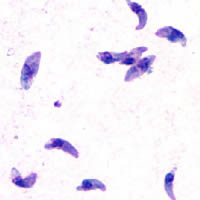
Los quistes tisulares son ingeridos por el gato (por ejemplo, al alimentarse de un ratón infectado). Los quistes sobreviven el paso por el estómago del gato y los parásitos infectan las células epiteliales del intestino delgado en donde pasan por la reproducción sexual y la formación de ooquistes, que son liberados con las heces. Otros animales, incluyendo los humanos ingieren los ooquistes (al comer vegetales no lavados adecuadamente) o los quistes tisulares al comer carne cruda o cocida inapropiadamente. Los parásitos entran a los macrófagos de la pared intestinal para luego distriburse por la circulación sanguínea y el cuerpo entero.
Historia


Taquizoítos de Toxoplasma gondii teñidos con tinción de Giemsa, a partir de una muestra de líquido peritoneal de ratón.
En 1908, Nicole y Manceux demuestran la presencia del parásito en un roedor el Ctenodactylus gondii. 
Toxoplasmosis
Artículo principal: Toxoplasmosis
Las infecciones por T. gondii tienen la facultad de cambiar el comportamiento de ratas y ratones, haciendo que se acerquen, en vez de huir del olor de los gatos. Este efecto es de beneficio para el parásito, el cual puede reproducirse sexualmente si es ingerido por el gato.6 La infestación tiene una gran precisión, en el sentido de que no impacta los otros temores de la rata, tal como el temor de los espacios abiertos o del olor de alimentos desconocidos. Se ha especulado que el comportamiento humano puede igualmente verse afectado de alguno modo, y se han encontrado correlaciones entre las infecciones latente por Toxoplasma y varias características, tales como un aumento en comportamientos de alto riesgo, tales como una lentitud para reaccionar, sentimientos de inseguridad y neurosis.7

T.gondii es un parásito intracelular con un citoesqueleto probablemente especializado para la invasión de células que parasitar. En azul YFP-α-Tubulina, en amarillo mRFP-TgMORN1.

Tratamiento
Se recomienda el empleo de Pirimetamina y Sulfonaminas, la primera actúa sobre la síntesis del ácido fólico y la segunda sobre la síntesis del ácido paraaminobenzoico (sobre taquizoitos, no en quistes).
Para la prevencion de la toxoplasmosis congénita en mujeres embarazadas se recomienda la espiramicina, ya que es menos tóxica. Este medicamento evita que los taquizoitos pasen el lago placentario hacia el feto. Si el parásito ya ha atravesado la placenta ya no es eficaz, aunque parece tener beneficio disminuyendo la carga parasitaria y por lo tanto disminuyendo la severidad de los síntomas en algunos casos.8 En inmunodeficientes se recomienda la combinación de pirimetamina con sulfadiazina, pero dada la mayor posibilidad de los pacientes infectados con VIH de alergia a las sulfas, en ocasiones es necesario usar la combinación de pirimetamina con la clindamicina.
Cuadro clínico
La etapa aguda de las infestaciones por toxoplasmosis pueden ser asintomáticas, pero a menudo aparecen síntomas gripales que conllevan a estadios latentes. La infección latente es también, por lo general, asintomática, pero en personas inmunosuprimidas (pacientes trasplantados o con ciertas infecciones), pueden mostrar síntomas, notablemente encefalitis, que puede ser mortal.
Varía dependiendo en qué trimestre del embarazo se adquiera el parásito:
La toxoplasmosis en embarazadas es rara vez sintomática pero puede provocar: linfadenopatía, fiebre, mialgia, malestar general, entre otras.

History of penicillin

The discovery of penicillin is attributed to Scottish scientist and Nobel laureate Alexander Fleming in 1928.[15] He showed that, if Penicillium notatum were grown in the appropriate substrate, it would exude a substance with antibiotic properties, which he dubbed penicillin. This serendipitous observation began the modern era of antibiotic discovery. The development of penicillin for use as a medicine is attributed to the Australian Nobel laureate Howard Walter Florey together with the German Nobel laureate Ernst Chain and the English biochemist Norman Heatley.
However, several others reported the bacteriostatic effects of Penicillium earlier than Fleming. The use of bread with a blue mould (it is presumed, penicillium) as a means of treating suppurating wounds was a staple of folk medicine in Europe since the Middle Ages. The first published reference appears in the publication of the Royal Society in 1875, by John Tyndall.[16] Ernest Duchesne documented it in an 1897 paper, which was not accepted by the Institut Pasteur because of his youth. In March 2000, doctors at the San Juan de Dios Hospital in San José, Costa Rica, published the manuscripts of the Costa Rican scientist and medical doctor Clodomiro (Clorito) Picado Twight (1887–1944). They reported Picado's observations on the inhibitory actions of fungi of the genus Penicillium between 1915 and 1927. Picado reported his discovery to the Paris Academy of Sciences, yet did not patent it, even though his investigations started years before Fleming's. Joseph Lister was experimenting with penicillum in 1871 for his Aseptic surgery. He found that it weakened the microbes but then he dismissed the fungi.
These early investigations did not lead to the use of antibiotics to treat infection because they took place in obscure circumstances, and the idea that infections were caused by transmissible agents was not widely accepted at the time. Sterilization measures had been shown to limit the outbreak and spread of disease; however, the mechanism of transmission of disease by parasites, bacteria, viruses and other agents was unknown. In the late 19th century, there was increasing knowledge of the mechanisms by which living organisms become infected, how they manage infection once it has begun and, most importantly in the case of penicillin, the effect that natural and man-made agents could have on the progress of infection.
Fleming recounted that the date of his discovery of penicillin was on the morning of Friday, September 28, 1928.[17] It was a fortuitous accident: in his laboratory in the basement of St. Mary's Hospital in London (now part of Imperial College), Fleming noticed a petri dish containing Staphylococcus plate culture he had mistakenly left open, which was contaminated by blue-green mould, which had formed a visible growth. There was a halo of inhibited bacterial growth around the mould. Fleming concluded that the mould was releasing a substance that was repressing the growth and lysing the bacteria. He grew a pure culture and discovered that it was a Penicillium mould, now known to be Penicillium notatum. Charles Thom, an American specialist working at the U.S. Department of Agriculture, was the acknowledged expert, and Fleming referred the matter to him. Fleming coined the term "penicillin" to describe the filtrate of a broth culture of the Penicillium mould. Even in these early stages, penicillin was found to be most effective against Gram-positive bacteria, and ineffective against Gram-negative organisms and fungi. He expressed initial optimism that penicillin would be a useful disinfectant, being highly potent with minimal toxicity compared to antiseptics of the day, and noted its laboratory value in the isolation of "Bacillus influenzae" (now Haemophilus influenzae).[18] After further experiments, Fleming was convinced that penicillin could not last long enough in the human body to kill pathogenic bacteria, and stopped studying it after 1931. He restarted clinical trials in 1934, and continued to try to get someone to purify it until 1940.[19]
"Discovery of penicillin"
 
. American Chemical Society.

Ántibacteriales - Antibióticos

Lista de antibacteriales
List of antibiotics

This poster from the U.S. Centers for Disease Control and Prevention "Get Smart" campaign, intended for use in doctor's offices and other healthcare facilities, warns that antibiotics do not work for viral illnesses such as the common cold.

Timeline of anti-infective therapy
This is the timeline of antimicrobial therapy. The years show when given was released onto the pharmaceutical market. Please note that this is NOT a timeline of the antibiotic itself!
An antibacterial is a compound or substance that kills or slows down the growth of bacteria.[1] The term is often used synonymously with the term antibiotic(s); today, however, with increased knowledge of the causative agents of various infectious diseases, antibiotic(s) has come to denote a broader range of antimicrobial compounds, including anti-fungal and other compounds.[2]
The term "antibiotic" was coined by Selman Waksman in 1942 to describe any substance produced by a microorganism that is antagonistic to the growth of other microorganisms in high dilution.[3] This definition excluded substances that kill bacteria but are not produced by microorganisms (such as gastric juices and hydrogen peroxide). It also excluded synthetic antibacterial compounds such as the sulfonamides. Many antibacterial compounds are relatively small molecules with a molecular weight of less than 2000 atomic mass units.
With advances in medicinal chemistry, most of today's antibacterials chemically are semisynthetic modifications of various natural compounds.[4] These include, for example, the beta-lactam antibacterials, which include the penicillins (produced by fungi in the genus 'Penicillium'), the cephalosporins, and the carbapenems. Compounds that are still isolated from living organisms are the aminoglycosides, whereas other antibacterials—for example, the sulfonamides, the quinolones, and the oxazolidinones—are produced solely by chemical synthesis. Accordingly, many antibacterial compounds are classified on the basis of chemical/biosynthetic origin into natural, semisynthetic, and synthetic. Another classification system is based on biological activity; in this classification antibacterials are divided into two broad groups according to their biological effect on microorganisms: bactericidal agents kill bacteria, and bacteriostatic agents slow down or stall bacterial growth.
History

Penicillin, the first natural antibiotic discovered by Alexander Fleming in 1928.
Before the early twentieth century, treatments for infections were based primarily on medicinal folklore. Mixtures with antimicrobial properties that were used in treatments of infections were described over 2000 years ago.[5] Many ancient cultures, including the ancient Egyptians and ancient Greeks used specially selected mold and plant materials and extracts to treat infections.[6][7] More recent observations made in the laboratory of antibiosis between micro-organisms led to the discovery of natural antibacterials produced by microorganisms. Louis Pasteur observed that, "if we could intervene in the antagonism observed between some bacteria, it would offer perhaps the greatest hopes for therapeutics".[8]
The term antibiosis, meaning "against life," was introduced by the French bacteriologist Vuillemin as a descriptive name of the phenomenon exhibited by these early antibacterial drugs.[9][10] Antibiosis was first described in 1877 in bacteria when Louis Pasteur and Robert Koch observed that an airborne bacillus could inhibit the growth of Bacillus anthracis.[11] These drugs were later renamed antibiotics by Selman Waksman, an American microbiologist in 1942.[3][9]
Antagonistic activities by fungi against bacteria were first described in England by John Tyndall in 1875.[8] Synthetic antibiotic chemotherapy as a science and development of antibacterials began in Germany with Paul Ehrlich in the late 1880s.[9] Ehrlich noted that certain dyes would color human, animal, or bacterial cells, while others did not. He then proposed the idea that it might be possible to create chemicals that would act as a selective drug that would bind to and kill bacteria without harming the human host. After screening hundreds of dyes against various organisms, he discovered a medicinally useful drug, the synthetic antibacterial Salvarsan.[9][12][13] In 1928, Alexander Fleming observed antibiosis against bacteria by a fungus of the genus 'Penicillium'. Fleming postulated that the effect was mediated by an antibacterial compound named penicillin and that its antibacterial properties could be exploited for chemotherapy. He initially characterized some of its biological properties, but he did not pursue its further development.[14][15] Prontosil, the first commercially available antibacterial antibiotic, was developed by a research team led by Gerhard Domagk in 1932 (who received the 1939 Nobel Prize for Medicine for his efforts) at the Bayer Laboratories of the IG Farben conglomerate in Germany.[13] Prontosil had a relatively broad effect against Gram-positive cocci but not against enterobacteria. The discovery and development of this first sulfonamide drug opened the era of antibacterial antibiotics. In 1939, Rene Dubos reported discovery of the first naturally derived antibiotic, gramicidin from B. brevis. It was one of the first commercially manufactured antibiotics in use during World War II to prove highly effective in treating wounds and ulcers.[16]
Florey and Chain succeeded in purifying penicillin. Purified penicillin displayed potent antibacterial activity against a wide range of bacteria and had low toxicity in humans. Furthermore, its activity was not inhibited by biological constituents such as pus, unlike the synthetic sulfonamides. The discovery of such a powerful antibiotic was unprecedented, and the development of penicillin led to renewed interest in the search for antibiotic compounds with similar efficacy and safety.[17] For their discovery and development of penicillin as a therapeutic drug, Ernst Chain, Howard Florey, and Alexander Fleming shared the 1945 Nobel Prize in Medicine. Florey credited Dubos with pioneering the approach of deliberately and systematically searching for antibacterial compounds, which had led to the discovery of gramicidin and had revived Florey's research in penicillin.[16]

Molecular targets of antibiotics on the bacteria cell
Like antibiotics, antibacterials are commonly classified based on their mechanism of action, chemical structure, or spectrum of activity. Most antibacterial antibiotics target bacterial functions or growth processes.[9] Antibiotics that target the bacterial cell wall (such as penicillins and cephalosporins), or cell membrane (for example, polymixins), or interfere with essential bacterial enzymes (such as quinolones and sulfonamides) have bactericidal activities. Those that target protein synthesis, such as the aminoglycosides, macrolides, and tetracyclines, are usually bacteriostatic.[28] Further categorization is based on their target specificity. "Narrow-spectrum" antibacterial antibiotics target specific types of bacteria, such as Gram-negative or Gram-positive bacteria, whereas broad-spectrum antibiotics affect a wide range of bacteria. Following a 40-year hiatus in discovering new classes of antibacterial compounds, three new classes of antibiotics have been brought into clinical use. These new antibacterials are cyclic lipopeptides (including daptomycin), glycylcyclines (e.g., tigecycline), and oxazolidinones (including linezolid).[29]